Laser beams
Optical techniques have recently discovered many new applications, ranging
from biology to the nuclear industry. These techniques provide information on
the chemical composition of the targets with spatial resolution down to
micrometers. The main advantages of optical techniques are fast and non-contact
measurements provided in real-time, and relatively low equipment costs.
The group of Optical micro-manipulation techniques offers the following
expertise:
- Construction of unique instrumentation for diagnostics that employs laser-based spectroscopy techniques such as Laser Induced Breakdown Spectroscopy (LIBS) and Raman spectroscopy.
- Applications of focused laser beams in micro-world for contactless manipulations and sorting of micro-objects, production of micro-structures using the technique of photopolymerization, and two- and three-dimensional modification of objects using laser ablation.
- Searching and development of new methods for identification of microorganisms, their separation or alternatively, destruction.
Applications of laser-induced breakdown spectroscopy – LIBS
Recently, there has been increased interest and demand in nearly all
scientific research areas for experimental techniques that enable fast and
unambiguous interpretation of the acquired data. This interest follows
industrial requirements for fast and accurate material identification and
analyses without lengthy laboratory techniques. The technique of LIBS involves
formation of luminous plasma generated by focusing the radiation from a pulsed
fixed-frequency laser (less then 1J/pulse) onto the surface of the sample to be
analysed. The light emitted by the hot plasma is dispersed by a spectrometer and
the characteristic emission lines produced by the excited elements during the
plasma cooling make it possible to determine the elemental composition of the
sample within the focus area. Detection limits of LIBS range from a few ppm to
hundreds of ppm (w/w).
The additional advantages and applications of LIBS:
- With LIBS the entire interaction between the system and the target is purely optical. Thus, the analysis can be performed remotely, provided that an optical connection can be established between the instrument and the target, e.g. with the use of an optical fibre. For remote applications the target can be placed up to hundreds of meters away from the mobile LIBS instrument (sediments inside storage tanks, oil rigs, and rigid steel constructions).
- LIBS excels in instances where on-site, real-time, and on-line measurements in various production phases are required (e.g. for metal identification problems). Different types of steel (e.g. FV520, NAG, 17/4, and 18/13) can be identified simply by following some of the representative elements (e.g. Mo, Ni, and Ti).
- LIBS can detect the following elements – Be, U, I, Al, C, Ca, Mg, Cr, Pb, Si, Li, Hg, Sr, Rb, Ti, Fe, Ni, V, Mn, Mo, etc.
- The most appropriate applications of LIBS are those encountered in the nuclear and chemical industry, where quantitative or qualitative remote analysis, without any physical contact with the sample, is preferred. A mobile LIBS setup based on the optical fibre delivery system can be identified as a natural solution to some of the analytical problems where exposure of personnel to highly radioactive or toxic material is understandably undesirable.
- Various setups can be used for environmental monitoring. Specially engineered systems can be designed and assembled for each analytical problem, allowing fast decisions to be made concerning the identification of target materials which can then be immediately sorted and labelled.
- LIBS can be used for quantitative analysis with reasonable precision even for small samples provided a tightly focused beam is used for material ablation (this is limited by the focusing optics).
Contact:
Mgr. Ota Samek, Dr.
e-mail: osamek@isibrno.cz
phone: + 420 541 514 127
Detailed information: http://www.isibrno.cz/omitec

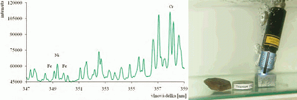
Emmision lines identified in the LIBS spectrum of a metal sample – Fe (left); LIBS setup for material identification of samples submerged in liquids (right)
Laser Raman spectroscopy
Laser Raman spectroscopy is a powerful technique for the identification of a
wide range of samples – solids, liquids, and gases. It is a straightforward,
non-destructive technique requiring no sample preparation. Raman spectroscopy
involves illuminating the sample with monochromatic light (laser) and using a
spectrometer to examine light that is scattered by the sample. Spectral
composition of the scattered light carries information about different molecular
or crystal vibrational modes which can serve as a unique characteristic for
different materials. This makes Raman spectroscopy a useful technique for fast
material identification (usually a few minutes are required) and
characterisation of the content of DNA, RNA, lipids, proteins, sugars, pigments,
saccharides, and amides within the sample. Typical Raman spectrometers for
material identification use a microscope to focus the laser beam on a small spot
so that the information about chemical composition from femtolitre volumes can
be obtained. Even though the principle of this method has been known for almost
a hundred years, only recently has started to be utilized in many unique
applications triggered especially by the development of sensitive detectors.
The advantages and applications of Raman spectroscopy
- Raman spectroscopy is capable of rapidly identifying biological samples. For example, medically relevant bacterial strains can be detected (e.g. Staphylococcus epidermidis). Raman spectroscopy can discriminate between biofilm-positive and biofilm-negative bacterial strains in real-time. Thus, time and cost of patient treatment related to virulent infection can be reduced because an appropriate therapeutic strategy can be chosen.
- Raman spectra can serve for identification of cells in oncological applications (e.g. diagnostics for early recognition of cancerous cells in-vivo and in-vitro) and identification of cells invaded by viral infections.
- Non-destructive analysis in the pharmaceutical industry (e.g. on-line characterization of tablets) and production of chemical maps of different surfaces or identification of nano-structures are possible.
- Raman spectroscopy can be combined with optical tweezers to form so-called Raman tweezers. This powerful combination can be used for identification and sorting of microorganisms freely moving in liquids.
Contact:
Mgr. Ota Samek, Dr.
e-mail: osamek@isibrno.cz
phone: + 420 541 514 127
Detailed information: http://www.isibrno.cz/omitec

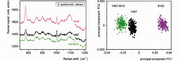
Typical Raman spectra of three bacterial strains of Staphylococcus epidermis (left); plot of Principle Component realition of three Staphylococcus epidermis strains (right)
Optical micro-manipulation techniques
Optical micro-manipulation techniques use the transfer of momentum from light
to microobjects. Such a transfer occurs, for example, during light scattering
from the microobjects, which is accompanied by a change of direction of the
light propagation. Optical micromanipulations make it possible to influence the
motion of objects of sizes from tens of nanometers to tens of micrometers just
by illumination with a laser beam. Optical tweezers represent an optical analogy
to the classical mechanical manipulation tool, using a single tightly focused
laser beam to confine objects in a contactless way. Since the objects are
trapped in the vicinity of the beam focus, repositioning of the focus also
causes repositioning of the objects, i.e. their controlled spatial
micro-manipulation. Several laser beam foci can be placed simultaneously within
the specimen in a controlled way, therefore enabling simultaneous confinement
and controlled micro-manipulation with multiple objects. This tool is mainly
used for manipulation of objects suspended in a liquid medium (living
microorganisms or cells in water or appropriate solution, microobjects placed
behind transparent obstacles etc.). Since such small objects can only be
observed using optical microscope, both techniques are often combined.
Examples of optical micromanipulation application:
- A compact version of optical tweezers has been developed based on an adapter inserted between the microscope and lens and containing an integrated laser diode or optical fibre connector. Optical micromanipulation is therefore possible without disturbing the optical path of the microscope.
- Optical tweezers can be combined with several optical spectroscopic techniques (e.g. Raman microspectroscopy, fluorescent spectroscopy) to enable contactless and non-destructive characterization of the trapped microobjects.
- Highly promising is the combination of optical micromanipulation techniques with microfluidic systems (lab-on-a-chip) that can serve, for example, for the study of stress at the level of individual cells followed by cell sorting.
- In addition to tightly focused beams there are also other configurations of laser beams for regular microobject confinement and arrangement in the space or on the surface.
- Illumination of moving microobjects influences their motion and can cause rectification of their stochastic motion leading to optical sorting of different suspension components (e.g. different types of cells).
Examples of applications of laser beams focused down to micrometer spots
- The significant increase of the laser beam intensity near its focus initiates photopolymerization – a chemical reaction that solidifies the liquid monomer into a solid polymer. Complex microstructures can be created by controlled positioning of the focused laser beam within the monomer solution.
- Focused pulsed laser beams of suitable wavelength offer many opportunities of employing their destructive effects (microablation) to volume or surface modifications of objects including, for example, intervention inside living cells.
Contact:
prof. RNDr. Pavel Zemánek, Ph.D.
email: zemanek@isibrno.cz
phone: +420 541 514 202
Detailed information: http://www.isibrno.cz/omitec

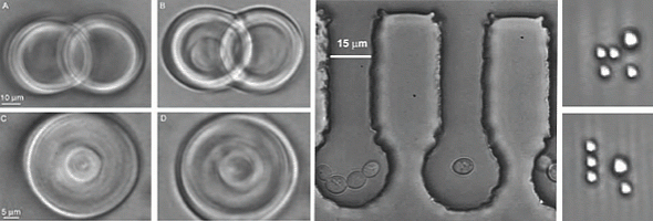
Examples of optical sorting of microobjects


