Measurement and data processing in medicine
The MediSIG team of the ISI provides a complete solution for neurology, cardiology and medical signal analysis and other applications.
The activities of the team include the development of new non-invasive
diagnostic procedures in cardiology and neurology, measurement and signal
processing in medicine and the design and construction of experimental medical
devices and acquisition systems. The MediSIG team cooperates with domestic and
foreign hospitals and physicians. Cooperation includes the implementation of
measurement and diagnostics protocols, design of recording parameters and
physiological signals, data digitizing and archiving, processing and detection
of physiological parameters and statistical analysis and evaluation of results.
The outcomes of interdisciplinary cooperation also include joint patents and
original experimental equipment and methodology.
MediSIG Cardiology – non-invasive diagnostics of cardiovascular
disease
Diagnostics is focused on determining the level of risk of coronary heart
disease, sudden cardiac death, arrhythmia, heart attack and heart failure, as
well as ischemic disease, high blood pressure risks and heart transplant
innervations. It is based on the evaluation of multi-lead ECG signal, blood
pressure, impedance cardiography, heart sounds and respiration. Results include
the determination of complete hemodynamic parameters, blood vessel stiffness and
control properties of blood circulation.
MediSIG Neurology – EEG signal processing, epilepsy,
Parkinson’s disease
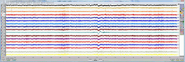
MediSIG provides complete processing, analysis and statistical evaluation of EEG recordings from deep brain structures. Epilepsy that cannot be pharmacologically controlled can be treated only with surgery. Intracerebral or subdural electrodes are implanted to localize epileptic sources. Unique EEG recordings from depth electrodes allow the development of new techniques for analyzing evoked processes, synchronization and desynchronization, and signal propagation directions and connectivity between different brain structures during various mental activities.
Software – methodology & processing
ScopeWin – system for measurement and data processing in medicine
Basic features:
- Data acquisition – ScopeWin communicates with the NIDAQMx library, which covers the complete series of National Instruments hardware.
- Visualization – ScopeWin includes a graphic editor that allows comfortable data inspection, signal comparison, manipulation, cursor operation etc. Data can be converted to conventional formats.
- Post-acquisition processing includes the following features: frequency analysis, digital filtering, functions for mathematical signal processing (splines, derivation, integration, digital quadrature detection and demodulation, regression, 50Hz signal detection, processing and elimination), two-dimensional analysis (time-frequency analysis – amplitude, phase and power), segmentation and averaging according to established criteria and several more. Most measurement, visualization and processing functions can be implemented as commands in the program and carried out sequentially without operator intervention.
ScopeWin signal processing in cardiology includes the following functions: ECG signal parameters detection (R wave, QT interval, heart rate, QT dispersion, T wave alternans), continuous blood pressure recordings analysis (systolic, diastolic, mean pressure, blood pressure variability, baroreflex), heart sounds (S1, S2), impedance cardiography (stroke volume and cardiac output, elasticity of blood vessels, pulse pressure wave) and respiration (breathing frequency and depth). ScopeWin provides procedures for complete evaluation of hemodynamic parameters. It also allows the implementation of new procedures and methods.
ScopeWin signal processing in neurology includes the following features:
visualization of large datasets of intracranial EEG recordings, time-frequency
analysis, segmentation, automatic removal of artificial segments and channels,
averaging and evoked potential calculation, envelope analysis, etc.
EEG signal analysis MediSIG provides professional advice with
the realization of EEG measurement and complex data analysis. We use our own
software (ScopeWin, ScopeMAT) equipped with advanced methods of analysis. The
software is intended for off-line data processing, especially for EEG, EKG, EMG
and EOG signals analysis. Processed data are obtained from clinical and
experimental intracerebral or scalp EEG recordings. Today they are recorded from
up to 128 recording points with a sampling frequency of 1kHz.
Data analysis includes:
- analysis of non-repeated signals (data recorded without subject stimulation);
- analysis of repeated events (data recorded during subject stimulation: visual/sound/tactile/etc.);
- impact of Deep Brain Stimulation (DBS) on EEG development.
Developed software packages cover complex off-line methods suitable especially for neurological data analysis. Our software products allow:
- data blocks manipulation, data preprocessing, data analysis based on FFT, visual data representation and basic statistical data parameterization;
- statistical evaluation and automation of data processes (ScopeMAT);
- analysis of connections between brain structures based on EEG data from intracerebral electrodes;
- visualization of results on a stereotactic brain model;
- complex software for analysis and visualization – EEGVisual.
ScopeMAT software allows parametric batch data processing covering: FFT
filtration, power analysis, time-frequency analysis, statistical evaluation of
repeated events (Wilkoxon test, t-test, bootstrap test), automatic detection of
power increase and decrease, automatic detection of statistically significant
components in time or frequent domain, statistical tests for determination of
repeated evoked signal differences recorded during different external conditions
(subject stimulation of type A/B/etc., test for two modalities, Schoffe test for
more than two modalities). Groups of subjects can also be evaluated using
ScopeMAT software. We can compute „GrandAverages“ and present significant
results for different contact groups transparently. The ScopeMAT software is
proposed for the MATLAB environment. This solution allows quick algorithm
implementation depending on application demand. The ScopeMAT software provides
numerical and graphical outputs to many format types, including MS Word and MS
Excel.
Analysis of EEG from depth electrodes, in cooperation with Brno Epilepsy center, Departments of neurology and neurosurgery St. Anne's University Hospital and Medical faculty of Masaryk University Brno, Czech Republic
Data acquisition systems
In addition to software and experimental devices MediSIG also produces
complete acquisition systems. It can simultaneously record up to 128 channels,
sampling up to 5MHz per channel with 16bit resolution and higher. The hardware
is built on top of National Instruments PCI / PCMCIA / USB devices. For
experiment control and data recording the ScopeWin program is used.
Realization examples:
- ISI Brno acquisition system.
- M&I – ISI high speed EEG recorder and analyzer (up to 200 channels, 40 kHz sampling each),
- ANNALab – acquisition system for neurology and cardiology, St. Anne’s University Hospital Brno, Mayo Clinic, USA
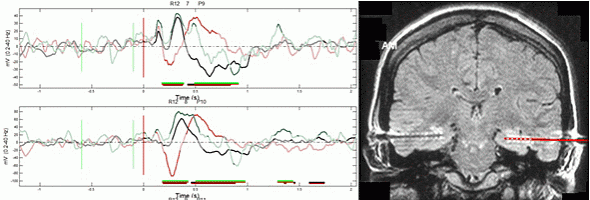
Intracranial EEG signal analysis, in cooperation with St. Anne's University Hospital Brno, Czech Republic
Experimental devices for measurement of biomedical signals
The MediSIG team mainly designs and constructs experimental devices for the
measuring and data processing of biological signals. We concentrate on devices
which are not available or do not yet have the desired parameters on the
commercial market.
Examples of devices:
- The Detector of Vagus Nerve Stimulation. The device for detection of activity of vagus nerve stimulation is intended for independent monitoring of implanted vagus nerve stimulation. It measures the electrical potential from the patient's neck, processes it and detects activity of the stimulator.
- RECO – device for respiration control. LEDs bargraph for pacing of breathing. It is used for controlled excitation of the cardiovascular system. It enables setting of pacing frequency 1–39 min-1, various modulations – ramp and sinus with depth of 25–100%.
- The Strain Gauge Plethysmograph. The strain gauge plethysmograph, which can be used for several measurement techniques in medicine, essentially measures the changes in human limb volume directly. However, it can be also used in indirect measurement for evaluating limb blood flow, blood pressure, digital volume pulses, for measuring circumferential changes of exposed arteries and breathing. Our device can measure up to 10 channels simultaneously.
- Experimental Syringe Mixing Pump For Ultrasound Contrast Agents. The pump enables continual infusion and bolus while mixing the contents of the syringe. The control of the pump is ensured by microcontrollers and is fully programmable by the user. The device is battery operated and has a robust mechanical construction. Technical parameters: syringe 10 ml and 20 ml type, infusion flow rate 0.3..4.0 ml/min, bolus flow rate 0 .3..4.0 ml/min, bolus time max. 60 seconds, mixing rate 30 cycles/min ±180º, minimum continuous working time 4.5 hours per one full battery charge.
- Multi-Channel Impedance Cardiography. Impedance
cardiography is a method based on measuring changes of thorax electrical
impedance. From the changes of thorax electrical impedance it is possible to
calculate important parameters of the cardiovascular system, like stroke volume
or cardiac output. The monitor makes it possible to measure small changes of
impedance on separate parts of the human body and to evaluate blood flow,
particularly the propagation of pulse waves in the whole body. It also makes it
possible to analyze vessel compliance. Commercial devices are equipped with one
or two channels and the simultaneous survey of blood flow in many parts of the
body is not possible. The main advantages of the monitor are the high quality of
the impedance signal, coherent impedance detection in all channels, measurement
of impedance phase changes and the option of stand-alone measurement without
computer and supply voltage.
- The multi-channel device consists of two-channel units. Each unit is comprised of two current generators and two voltage detectors. The device makes it possible to interconnect up to 9 units with clock and synchronization signals and to build an 18-channel device. The unit stores the impedance signal on a SD/SDHC memory card, transfers it in real-time to the PC through the USB port, and possibly represents it on analogue output.
- The parameters of the unit are:
- measurement frequency 30 to 70 kHz or 1 kHz to 1 MHz (optional)
- measurement current 1 mA, adjustable
- clock frequency of direct digital synthesis, AD converter and synchronous detector 60 MHz
- overall dynamic range of 127 dBc/√Hz
- phase resolution better than 0.001 degree
- output data format 16-bit complex envelope at 10 kHz sampling
- operating time 3 hours
- Device for microneurography. The dedicated preamplifier and amplifier with signal filtering and detection are used for monitoring the electrical activity in the nerves. The amplifies and processed signals obtained by means of microelectrodes (special insulated needle with conductive tip) directly from the desired part of nerves.
- ECG 12. Experimental 12 lead electrocardiograph amplifier with broad pass band up to 500 Hz. This device is intended to be used for special ECG measurements that cannot be performed with a standard commercial device. The amplifier is battery operated and is equipped with the 12 analog outputs, 1 monitor output and USB data acquisition unit. Figure PowerPoint 6
MediSIG has also designed and constructed a wide range of various devices for
measuring biological signals like electrical amplifiers and filters, pressure
measurement devices for the monitoring of breathing, pressure monitoring in
lungs and esophagus, devices for phonocardiography – special microphones with
dedicated amplifiers, spirometers, etc. The majority of these devices are
battery operated to enable use during measurements of weak biological signals,
where using AC mains operated devices cause unacceptable artifacts in the
measured signal.
Contact:
Dr. Pavel Jurák
email: jurak@isibrno.cz
phone: +420 541 514 312
Detailed information: http://www.isibrno.cz
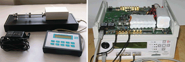
Experimental Syringe Mixing Pump for Ultrasound Contast agents (left), Multi-Channel Impedance Cardiography (right)