Nuclear magnetic resonance
The research of the Nuclear Magnetic Resonance group is oriented primarily
towards methodology of localized in-vivo MR spectroscopy, spectroscopic imaging,
and quantitative imaging such as diffusometry and relaxometry. These
nondestructive, noninvasive and non-ionising measurements are based on the
nuclide-selective interaction between magnetic fields produced by the NMR
scanner with the atomic nuclei located in the sample volume. The highest
sensitivity is achieved with 1H nuclei (protons) and, therefore, the prominent
targets are water molecules, which are abundant in biological samples and many
materials. In these measurements, the nuclei observed may not only describe the
anatomy of the sample interior, but they also act as atomic probes reporting on
the nearest environment and processes occurring on the time scale of picoseconds
to hours. Besides the ability to identify certain molecules, the acquired data
can provide a statistical representation of the fast and random atomic and
molecular interactions (manifested by relaxation times, diffusion coefficients,
chemical exchange rates and seen as intensity modifications) as well as
temporally and/or spatially resolved information on much slower processes
(concentration changes, perfusion, flow or motion seen in consecutive images).
Quantitative images mapping the spatial distribution of certain well-defined
physical properties (relaxation times, diffusion coefficients and directions,
flow velocity, temperature etc.) may be obtained and the presence of
paramagnetic or superparamagnetic nanoparticles can be detected.
Equipment:
With the MR scanner(s) available in ALISI, virtually all kinds of measurements
known from clinical MR scanners can be performed. Thanks to the increased
sensitivity resulting from the high magnetic field and small coils, the
achievable spatial resolution is much more adequate to the study of small
samples. The application sphere is not limited to biomedical imaging; techniques
targeting specific problems may be developed.
- MR scanner 4.7 T / 210 mm (proton resonance frequency 200 MHz), optimum sample diameter 40 mm, maximum diameter 100 mm, maximum length about 300 mm; measurable nuclei: 1H, 13C, 19F, 31P, 23Na, 129Xe (immediately), other nuclei negotiable (depending on the availability of RF coils and filters).
- MR scanner 9.4 T / 210 mm (proton resonance frequency 400 MHz, as of mid 2011).
- Physiological functions monitor and isoflurane gas anesthesia unit for small animals (as of 2011).
- RF laboratory.
Imaging and data analysis
ALISI is prepared to optimize the measurement and data processing protocols
for answering specific questions and to carry out the necessary measurements.
Due to the complexity of the technology and the enormous flexibility of NMR
measurements, collaborative work may be the most suitable way to maximizing the
synergy between the core competence of the client and the NMR competence of the
ALISI teams.
MR imaging, localized spectroscopy and spectroscopic
imaging
- Spatial mapping of 2D slices or 3D volumes, spatial resolution down to about 0.1×0.1×1 mm3 (10 nL) now, 0.05×0.05×1 mm3 (2.5 nL) as of 2011.
- Images weighted by spin density, T1, T2, T2*, diffusion, magnetic susceptibility.
- Molecule-specific images (water, fat, specific small molecules).
- Quantitative imaging of relaxation, diffusion, perfusion, flow, temperature changes, magnetic susceptibility.
- Mapping of the 3D spatial distribution of selected small molecules, particularly for in vivo studies of cell cultures, plants, extracted tissues, small animals (mouse, rat – as of 2011).
- Quantization of metabolite ratios in vivo.
MR data analysis
- MR imaging and spectroscopic imaging: own software Marevisi (developed in cooperation with National Research Council Canada – Institute for Biodiagnostics) – processing and visualization of multidimensional MR data sets.
- MR spectroscopic imaging: quantum-mechanics based simulations and data processing, software jMRUI (developed in collaboration by a consortium of European research institutions).
- Customized MR data analysis solutions.
- Evaluation of perfusion studies.
Education and training
- Customized courses, demonstrations and system-operation training available to pre- or postgraduate university programs or research-oriented professionals.
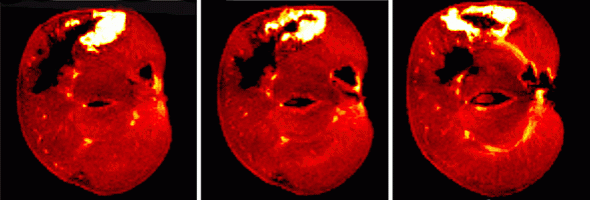
Slices of a decomposting worm-eaten apple (the hyperintense area results from faster relaxation of the smaller moleculs of the decomposed tissue
Application fields
Biomedical research
Numerous applications of the small-bore high-field MR scanner measurements are possible in the fast growing areas of nanomedicine, in the development and testing of
- stem cell based therapies for cardiology, neurology, orthopedics, diabetes treatment;
- targeted drugs for oncology, cardiology, neurology, psychiatry;
- contrast agents based on superparamagnetic nanoparticles.
High spatial resolution measurements of cell cultures, soft tissues,
cartilage, excised organs, or in-vivo in mice or rats may be the bridge
translating the molecular and cellular techniques to human medicine. It is an
advantage that the same NMR techniques can be applied in models and in humans.
Therefore, collaboration in human-oriented MR-based research is also possible,
particularly in areas matching the team’s research orientation. Biomedical
molecular imaging, combining NMR with optical modalities (fluorescence,
bioluminiscence, and/or Raman spectroscopy) is promising for the future.
The NMR expertise of ALISI may be further applied in other areas related to the
biomedical sector, such as
- design and construction of NMR phantoms and coils for special purposes;
- testing of MR compatibility of materials and implants;
- study of plant physiology.
Industrial technology NMR imaging and/or spectroscopy may be applied in various areas of the chemical, food, building, energy-producing industries, such as
- characterization of porous materials and gels by relaxometric and diffusometric methods;
- measurement of magnetic susceptibility of materials;
- development of biotechnologies.
Contact:
Dr. Zenon Starčuk jr.
email: starcuk@isibrno.cz
phone: +420 541 514 247
Detailed information: http://www.isibrno.cz
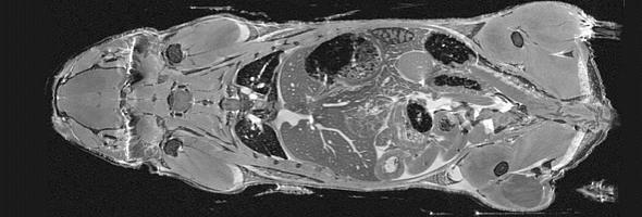
Mouse imaged in a 9.4 T MR scanner